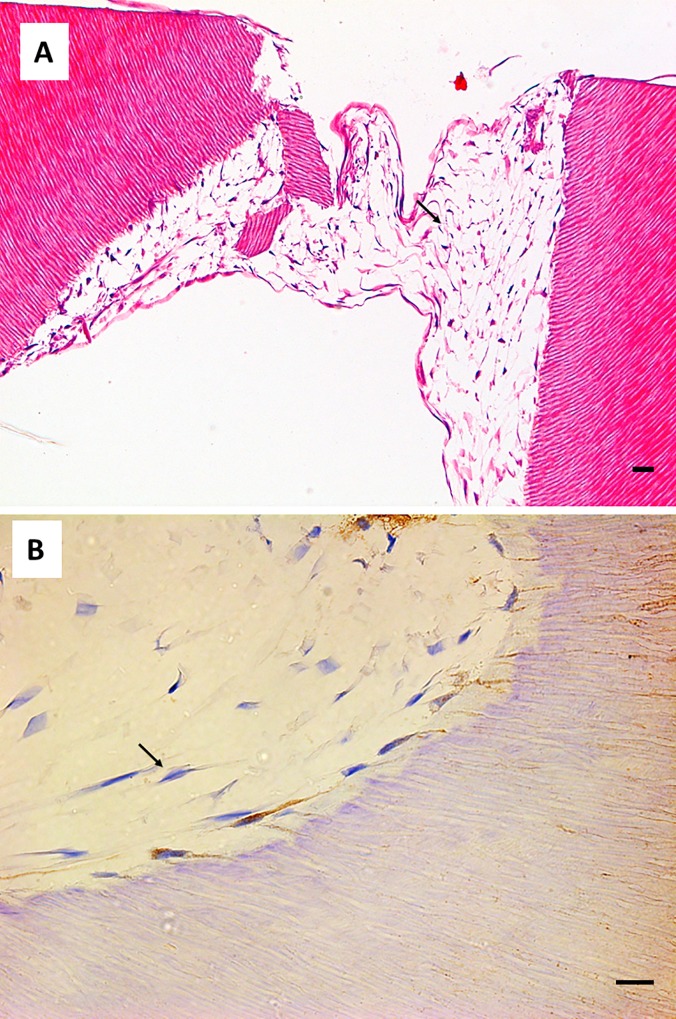
Fig 8. Cell morphology of hDPSCs cultured on SA.
hDPSCs were cultured on scaffolds for 6 weeks and processed for light microscopy. Samples were embedded in paraffin, cut into sections, and subjected to hematoxylin and eosin staining (panel A) or immunohistochemistry for DSPP (panel B). Black arrows indicate stellate cells negative for DSPP. Representative results of 12 separate experiments are shown. Scale bar equals to 20 μm.

