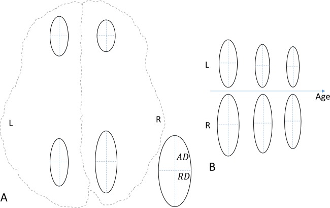
Fig 5. Schematic diagrams of the microstructural asymmetry of simplified diffusion tensors represented by ellipses.
(A) The front brain shows leftward asymmetry, and the back brain shows rightward asymmetry. (B) Asymmetry increases with age in the back brain. AD, axial diffusivity; RD, radial diffusivity; L, left; R, right; A, anterior; P, posterior. These diagrams are based on summaries of the results shown in Figs 2–4.

