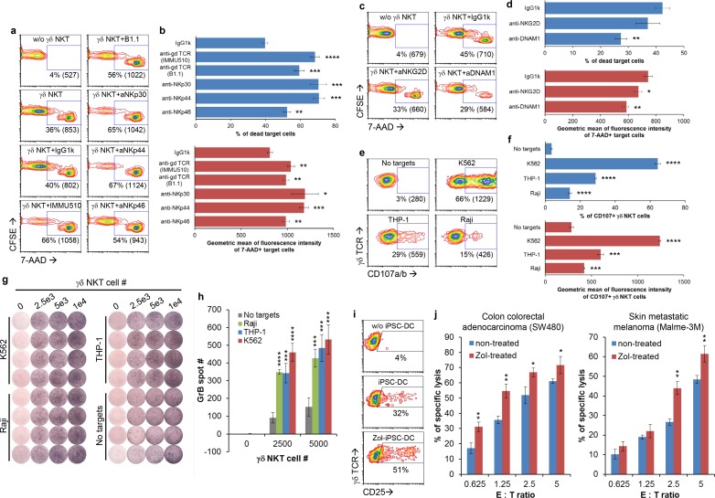
Fig 6. Mechanism of action of γδ NKT cell-mediated cytotoxicity.
(a-b) γδ NKT cells generated from GDTA/NF-iPSC#1 were used for redirected cytotoxicity against THP-1 cells in the presence of indicated activating antibodies. (c-d) Cytotoxicity of γδ NKT cells against THP-1 were studied in the presence of indicated blocking antibodies. Representative contour plots (a, c) and result summaries (b, d) were shown. The numbers in contour plots were % of 7-AAD+ THP-1 cells and geometric mean of fluorescence intensity of these cells (numbers in brackets). (e-f) CD107a/b expression on γδ NTK cells after coculture with the indicated cancer cells. Representative contour plots (e) and result summaries (f) were shown. The numbers in contour plots were % of CD107a/b+ γδ NKT cells and geometric mean of fluorescence intensity of these cells (numbers in brackets). (g-h) GrB secretion by γδ NKT cells upon coculture with the indicated cancer cells. ELISPOT images (g) and a summary of spot counting (h) were shown. (i) CD25 expression on γδ NKT cells after stimulation with syngeneic zoledronic acid-treated iPSC-DCs. (j) Cytotoxicity of γδ NKT cells against zoledronic acid-treated cancer cells. The statistical significance of differences while comparing with corresponding controls in above experiments was determined by Student’s t-test (mean ± SD, n = 3, *p < 0.05, **p < 0.01, ***p < 0.001, ****p < 0.0001).

