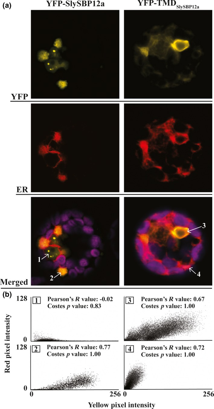
Figure 8.

Endoplasmic reticulum localization of SlySBP12a and TMDS ly SBP 12a in tomato protoplasts. Tomato protoplasts were transfected with plasmids encoding 35S:YFP‐SlySBP12a or 35S:YFP‐ TMDS ly SBP 12a and imaged by CLSM. An SP‐mCherry‐HDEL construct was cotransfected to serve as an ER marker (red). The magenta signal represents chloroplast autofluorescence. (a) Representative images of tomato protoplasts expressing 35S:YFP‐SlySBP12a or 35S: TMDS ly SBP 12a with the ER marker. Numbered regions indicated by arrows were used for colocalization analysis. (b) Intensity histograms of the four regions selected for colocalization analysis. Pearson's R values and Costes p‐values are displayed for each region
