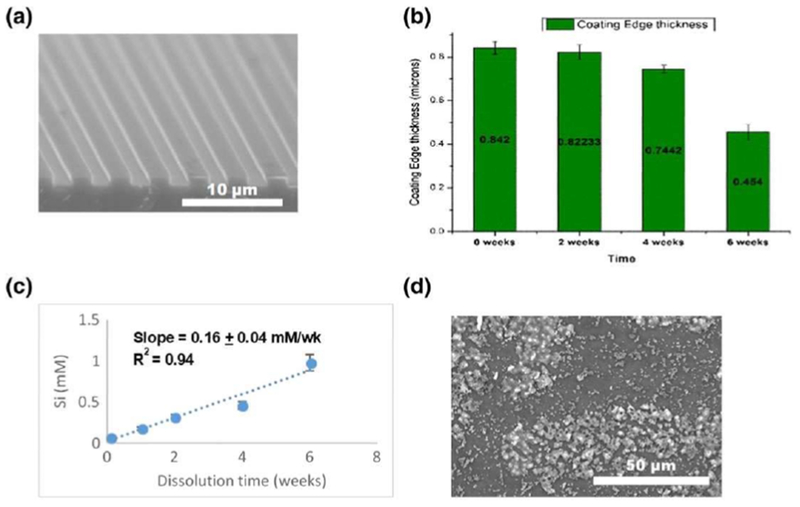
FIGURE 8.

SiONx coatings on implant surfaces (a) was immersed in alpha-MEM for 6 weeks. The coating was observed to degrade over 6 weeks (b) and estimated for the approximate rate of ionic Si release (c). The resultant surface appeared to have the formation of hydroxyapatite crystals in a c-axis orientation (d) [Colour figure can be viewed at wileyonlinelibrary.com]
