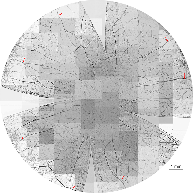
Figure 1.
Representative image of cat whole-mount corneal stromal nerves. The stromal nerves enter the cornea around the limbus as thick trunks and divide into many branches. These branches go toward the center and connect with each other to constitute the stromal nerve network. The entire cornea was labeled with PGP9.5, whole mount images were acquired using a fluorescent microscope with a 5x objective lens and images inverted to the white background. The picture was reconstructed from more than 100 images. The arrows indicate the main trunks of stromal nerves close to the limbus.

