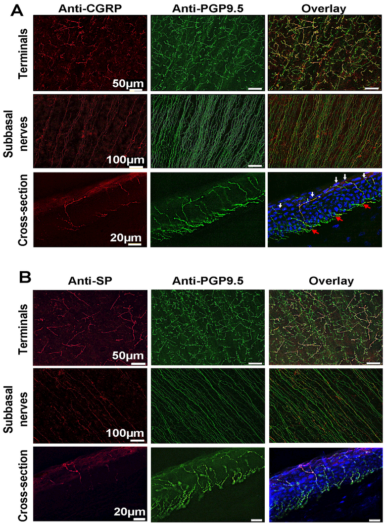
Figure 5.
Representative whole-mount and cross-section images in A) and B) show the expression of CGRP- and SP-positive nerves in the central subbasal bundles and terminals. In the cross-sections, DAPI (blue) was used to counterstain cell nuclei; red arrows show the subbasal nerves; white arrows indicate the terminals.

