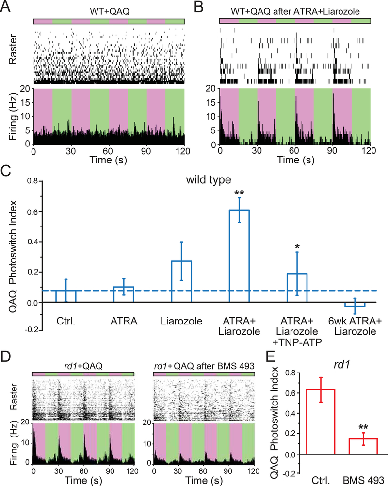
Figure 4. Pharmacological activation of RAR is necessary and sufficient for degeneration-dependent chemical photosensitization.
(A-B) MEA recordings from QAQ-treated WT retina, without (A) or with (B) prior intravitreal injection of ATRA plus liarozole. Photoswitching was elicited by alternating between 380 nm (purple) and 500 nm (green) light. 300 μM QAQ was applied onto the isolated retina for 30 minutes and then washed away.
(C) Quantification of (A-B). Photosensitivity (Photoswitch Index, PI) induced by QAQ was measured in WT retinas (blue). Recordings were obtained 3–7 days after ~1 μl intravitreal injection, including 1% DMSO in PBS (vehicle control, ‘Ctrl.’, dotted line in blue), 0.1 μM all-trans retinoic acid (ATRA), 100 μM Liarozole (Cyp26 inhibitor). Recordings were also obtained 6 weeks after injection of ATRA + Liarozole. 200 μM TNP-ATP (P2X antagonist) was bath loaded. ** - Ctrl. vs ATRA + Liarozole, * - ATRA + Liarozole vs ATRA + Liarozole + TNP-ATP.
(D) Blocking RAR reduces QAQ-photosensitization in rd1 retinas. Retinas were obtained from eyes without (left) or with (right) BMS 493 (0.5 μΜ), injected into the vitreous at 3–7 days prior to retina isolation and recording.
(E) Quantification of (D) in rd1 retinas (red). Control (‘Ctrl.’, non-injected eyes), were compared to rd1 eyes injected with 0.5 μΜ BMS 493, 3–7 days prior to recordings. (C&E) Values are shown as mean ± SEM. *p<0.05, **p<0.01, Kruskal-Wallis and t-test. For full datasets, see Table S1.

