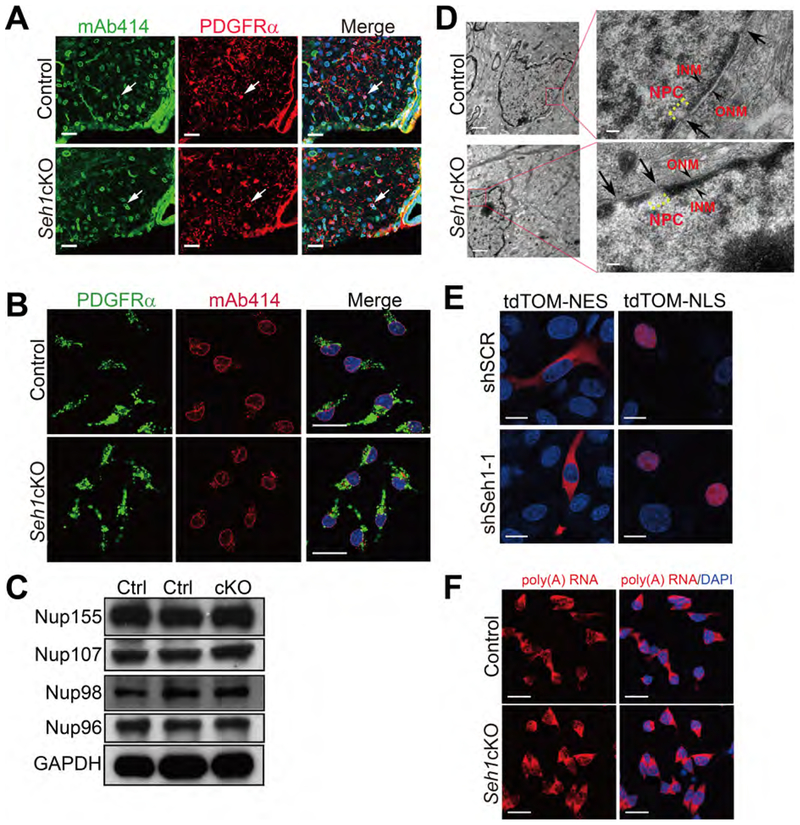
Figure 4. Ablation of Seh1 Does not Cause Major Defects in NPC Assembly or Function.
(A) Immunostaining of mAb414 and PDGFRα in spinal cord of P14 control and Seh1cKO mice. Arrows indicate mAb414+/PDGFRα+ cells. Nuclei were stained with DAPI. Scale bars=25 μm.
(B) Immunostaining of mAb414 and PDGFRα in primary control and Seh1cKO OPCs. Scale bars=25 μm.
(C) Immunoblot was performed to detect the indicated proteins in the spinal cord lysate of control and Seh1cKO at P14. Ctrl: control; cKO: Seh1cKO.
(D) Electron microscopy images of oligodendrocytes from control and Seh1cKO optic nerve. Boxed image is shown in the right. Arrows indicate NPCs. Arrowheads indicate ONM and INM. ONM/INM, outer/inner nuclear membrane. Scale bars=1 μm in left panel and 0.1 μm in right panel.
(E) Immunofluorescence of tdTomato-NES and tdTomato-NLS signals in Oli-neu cells transduced with scrambled or Seh1 shRNAs. NES, nuclear export signal; NLS, nuclear localization signal. Scale bars=15 μm.
(F) Oligo-dT In situ hybridization followed by fluorescence microscopy was performed in control and Seh1cKO OPCs. Scale bars=25 μm.
See also Figure S4.

