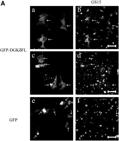
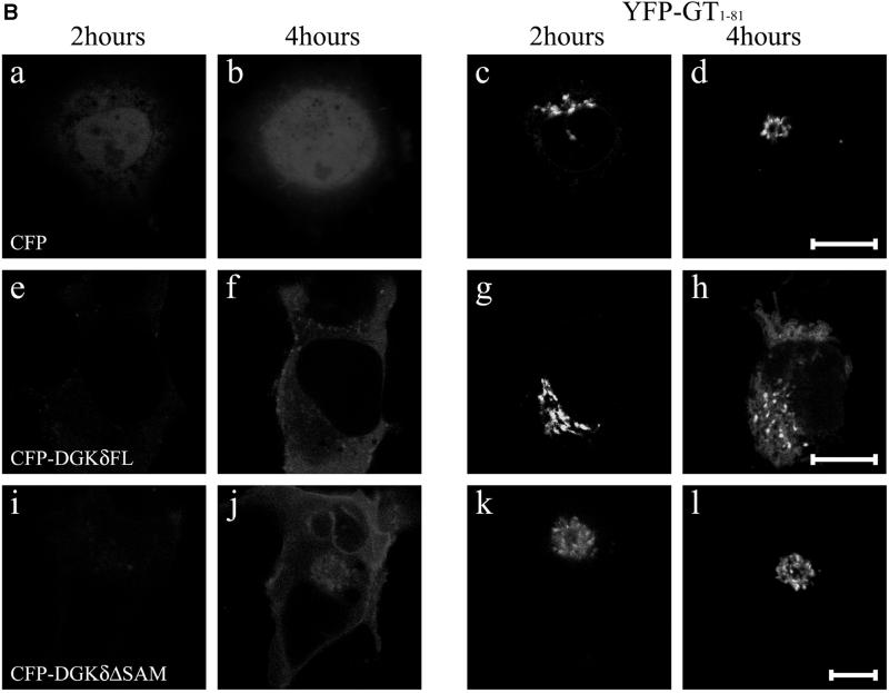
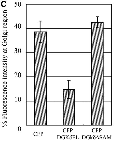
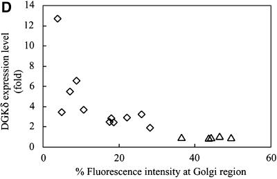
Figure 5.
Redistribution of Golgi membrane proteins caused by DGKδ expression. (A) COS7 cells were transfected with the expression plasmids for GFP-DGKδFL (a–d) or GFP (e and f) using Fugene 6. At 36 h after transfection, the cells were fixed with paraformaldehyde and immunostained with anti-GS15 antibody (Transduction Laboratories, Lexington, KY) and Alexa 594–conjugated anti-mouse antibody (b, d, and f). Arrows show cells expressing GFP-DGKδFL or GFP. Bars, 50 μm. (B) pEYFP-GT1–81 and expression vectors for CFP (a–d), CFP-DGKδFL (e–h), or CFP-DGKδΔSAM (i–l) were introduced into COS7 cells using siliconized glass microbeads as described in MATERIALS AND METHODS. Confocal images were taken after 1 h at 30-min intervals. Shown are the images at 2 h (a, c, e, g, i, and k) and 4 h (b, d, f, h, j, and l) of plasmid loading. Bars, 10 μm. (C) On the basis of the assumption that the juxtanuclear aggregates with intense signal of YFP-GT1–81 represented the Golgi region, we selected such areas in the LSM images by setting threshold fluorescence levels using IPLab software (Scanalytics, VA). Distribution of YFP-GT1–81 was calculated by dividing fluorescent intensities at the Golgi by those of whole cell signal at 4 h after plasmid loading. Mean values ± SEM of four cells are presented. (D) Expression levels of DGKδ and distribution of YFP-GT1–81. Either of the vectors for CFP (open triangles) or CFP-DGKδFL (open rhombuses) was cointroduced into COS7 cells with the vector for YFP-GT1–81 using microbeads. At 4 h after loading, the cells were fixed with methanol and immunostained with anti-DGKδ antibody or preimmune IgG followed by secondary anti-rabbit antibody conjugated to Alexa 594 (Molecular Probes, Eugene, OR). The YFP and Alexa594 signals of the LSM images were quantitated by IPLab. In quantitating the Alexa594 signal, the average signal obtained by the preimmune IgG incubation was set to zero. Distribution of YFP-GT1–81 was calculated as in C. The relative expression levels of DGKδ were obtained by dividing the whole-cell Alexa 594 signal in CFP-DGKδFL–expressing cells by the averaged endogenous DGK signal in control CFP-expressing cells and were compared with distribution of YFP-GT1–81 in the same cell.


