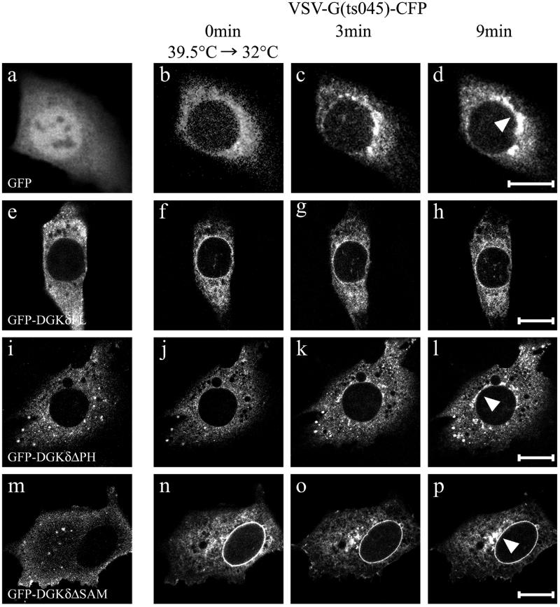
Figure 9.
DGKδ slowed the ER-to-Golgi transport of VSV G(ts045). Kinetics of ER-to-Golgi transport was measured in NIH3T3 cells expressing temperature-sensitive folding mutant, VSV G(ts045) fused to CFP (b–d, f–h, j–l, and n–p). The cells coexpressed GFP (a–d), GFP-DGKδFL (e–h), GFP-DGKδΔPH (i–l), and GFP-DGKδΔSAM (m–p). Cells transfected with the expression vectors were cultured first at nonpermissive temperature (39.5°C) for 24 h (=0 min; a, b, e, f, i, j, m, and n), and then the incubation temperature was shifted to permissive temperature (32°C). Thereafter, the confocal images were recorded at 1-min intervals. Images taken at 0 (a, b, e, f, i, j, m, and n), 3 (c, g, k, and o), and 9 min (d, h, l, and p) are shown. Arrowheads indicate the Golgi areas. Bars, 10 μm.

