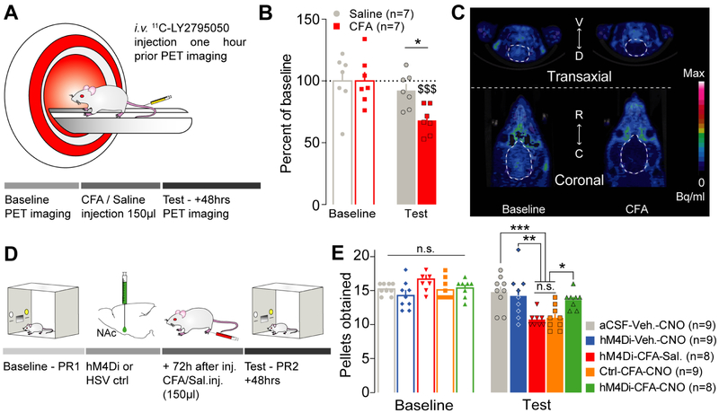
Figure 4: Inflammatory pain mediates negative affective states through recruitment of dynorphin-containing neurons in the NAcShCS.
(A) Schematic representation of the PET imaging methodology in rats. (B) Inflammatory pain, but not saline control, decreases the distribution volume of 11C-LY2795050 observed in rat’s brain two days after CFA injection (two-way ANOVA for repeated measures: time: F1,12=26.88, p=0.0002; interaction: F1,12=10.02, p=0.0081; post hoc tests: baseline CFA vs test CFA: p<0.0001; baseline saline vs test saline: p=0.3260; test CFA vs test saline: p=0.0282, n=7). (C) Representative images of PET imaging before (left) and after (right) inflammation. (D) Schematic representation of the behavioral methodology. (E) Silencing dynorphin neurons in the NAcShCS prevented the inflammatory pain-induced decrease in motivation for sucrose self-administration (two-way ANOVA for repeated measures: time: F1, 38= 75.81, p<0.0001; interaction: F4,38= 15.84, p<0.0001. Post-hoc test: aCSF-Veh.-CNO vs hM4Di-CFA-Sal.: p=0.0006; aCSF-Veh.-CNO vs Ctrl-CFA-CNO: p=0.0003; Ctrl-CFA-CNO vs. hM4Di-CFA-CNO: p=0.0255; hM4Di-CFA-Sal. vs hM4Di-CFA-CNO: p=0.0417; hM4Di-CFA-Sal. vs. hM4Di-Veh.-CNO: p=0.0046; Ctrl-CFA-CNO vs. hM4Di-Veh.-CNO: p= 0.0027; hM4Di-CFA-CNO vs. hM4Di-Veh.-CNO: p=0.9658; hM4Di-CFA-Sal. Vs Ctrl-CFA-CNO: p=0.9988; aCSF-Veh.-CNO vs. hM4Di-Veh.-CNO: p=0.9706; and aCSF-Veh.-CNO vs hM4Di-CFA-CNO: p=0.7173, n=8-9).

