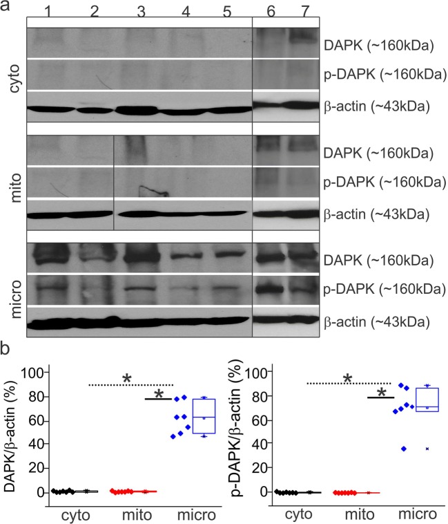
Fig. 2.
Subcellular localization of DAPK and p-DAPK in the epileptic brain. a Western blots of TLE brain specimens (#1–7) show that DAPK and p-DAPK are both localized in the microsomal (micro) fractions rather than to the cytoplasmic (cyto) or mitochondrial (mito) fraction (n = 7), indicating that DAPK activity is centered around the endoplasmic reticulum. b The relative ratio of DAPK/p-DAPK to β-actin increases only in the microfraction. Data is expressed as mean ± S.E.M. *p < 0.05. ANOVA, analysis of variance

