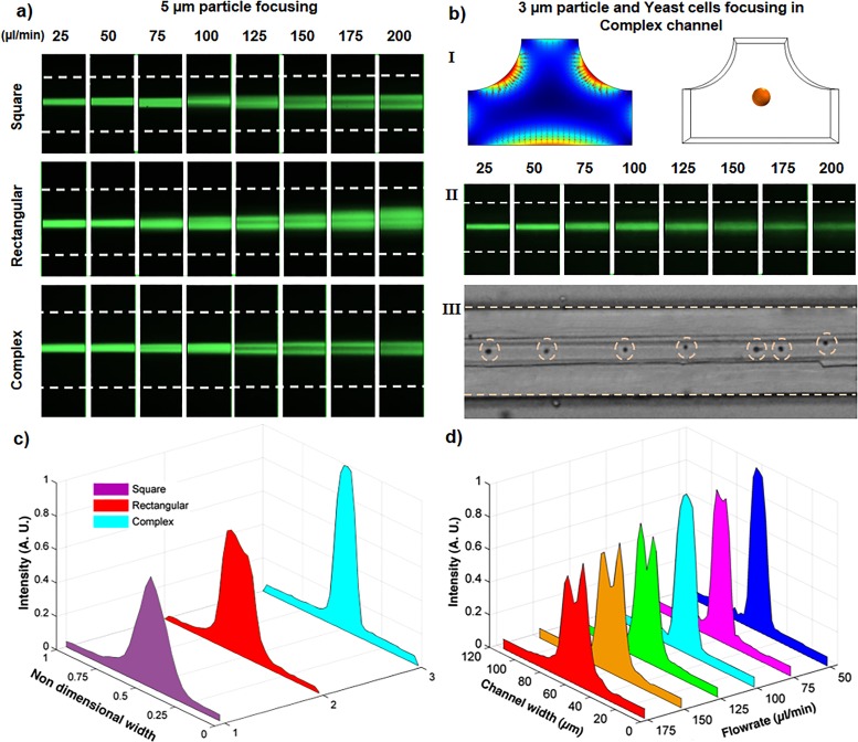
FIG. 8.
Focusing positions in square, rectangular, and complex channels. (a) Top view focusing positions of 5 μm particles, which shows for all the flow rates the complex channel has tighter focusing compared to the conventional channels. (bI) Elasto-inertial force vector plot in the modified complex channel and the focusing position for 3 μm particles. (bII) Top view focusing of the 3 μm fluorescent particles in the modified complex channel. (bIII) Focusing of the yeast cells in the complex channel for a flow rate of 75 μl/min. (c) Intensity profiles for the focusing of 5 μm particles at a flow rate of 100 μl/min, which shows a tighter focusing in the complex channel. (d) Intensity profiles for focusing of 5 μm particles through the complex channel.

