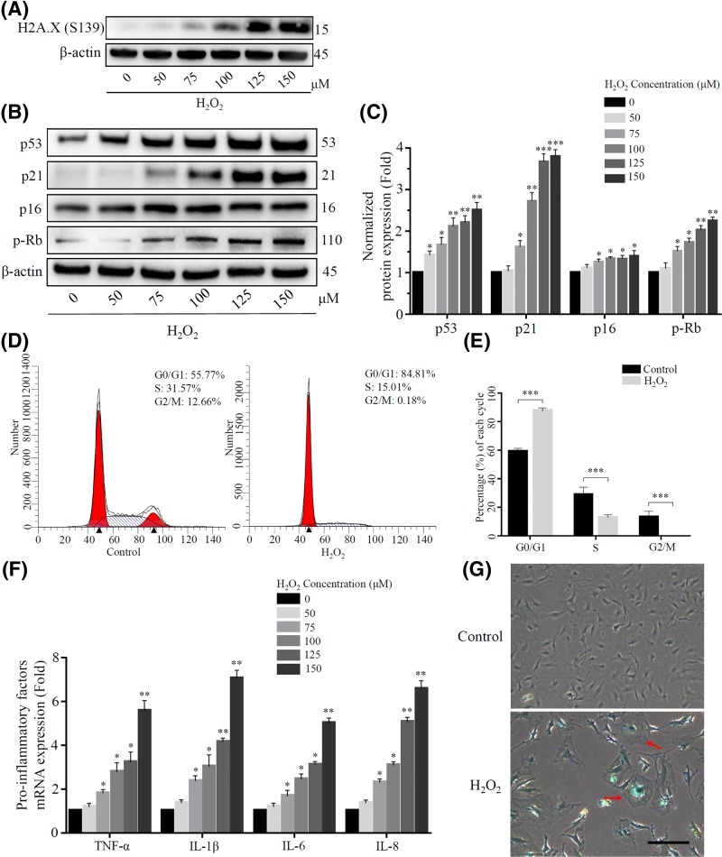
Figure 3. Sublethal concentration of H2O2 induced senescence in rat NP cells.
(A) DNA damage caused by H2O2 was reflected by the expression of Phospho-Histone H2A.X (Ser139). (B) and (C) The expression of some senescence relative proteins (p53, p21, p16 and p-Rb) were detected using Western blot after exposure to a long-term H2O2. β-actin was used as an internal control. (*P<0.05, **P<0.01, ***P<0.001 vs control group) (D) and (E) The cell cycle was detected by flow cytometry, and the proportion of cells in G0/G1 phase of H2O2 treatment group was higher than that of the control group. (***P<0.001 vs control group) (F) Some pro-inflammatory factors (TNF-α, IL-1β, IL-6 and IL-8) increased at the transcriptional level with the raising of H2O2 concentration. (*P<0.05, **P<0.01 vs control group) (G) The H2O2 treatment group showed more abnormal and β-gal-positive cells than the control group. Scale bars 100 μm.

