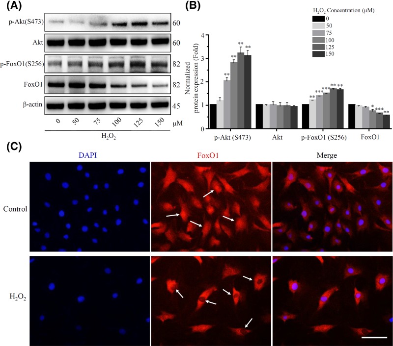
Figure 6. H2O2 increased FoxO1 phosphorylation by activating the PI3K/Akt pathway.
(A) and (B) The proteins expression of Akt, FoxO1 and their phosphorylation were detected using Western blot after exposure to a long-term H2O2. β-actin was used as an internal control. (*P<0.05, **P<0.01, ***P<0.001 vs control group) (C) Immunofluorescence staining was used to detect the expression and localization of FoxO1 in rat NP cells. The FoxO1 showed red fluorescence, and the nucleus showed blue fluorescence stained by DAPI. Scale bars 50 μm.

