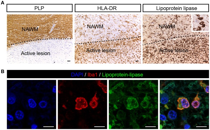
Figure 2.
LPL is expressed on iba1 positive cells in active MS lesion. (A) Active white matter lesion is characterized by loss of PLP. Active lesions showed enhanced LPL immunoreactivity in microglia/macrophages (insert) (scale bar = 50 μm). (B) Double immunofluorescent labeling shows presence of lipoprotein-lipase (green) positive Iba1 (red) cells in MS lesions (scale bar = 10 μm).

