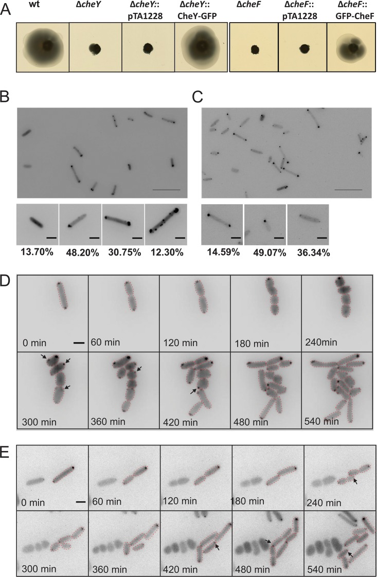
FIG 6.
Intracellular distribution and mobility of the CheY response regulator and the CheF adaptor protein in H. volcanii. (A) Influence of CheY and CheF on directional movement. The panels show results of assays of the motility of different H. volcanii strains on semisolid agar plates. pTA1228, empty plasmid. (B and C) Intracellular distribution of CheY-GFP in H. volcanii ΔcheY (B) and of GFP-CheF in H. volcanii ΔcheF (C) in the early log phase. The lower panels show closeup views of two different observed distribution patterns, and the numbers at the bottom represent percentages of the total population displaying the distribution (n, >1,000). Scale bars, 10 μm (upper panel) and 2 μm (lower panels). (D and E) Time-lapse images of dividing CheY-GFP-expressing ΔcheY H. volcanii cells (D) and of GFP-CheF expression in ΔcheF H. volcanii cells (E). Arrows indicate newly apparent foci. Scale bar, 2 μm. See also Movies S6 and S7 at https://doi.org/10.6084/m9.figshare.7718486 and https://doi.org/10.6084/m9.figshare.7718495.

