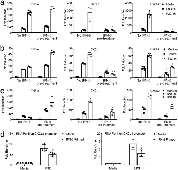
FIG 4.
IFN-β suppresses transcriptional induction of chemokine gene expression in vitro. (a) WT C57BL/6J macrophages were pretreated for 1 h with medium alone or IFN-β (100 U/ml). Macrophages were stimulated with the TLR2 agonist P3C (250 ng/ml) for 2 h or 4 h. RNA was harvested, and mRNA gene expression was analyzed by qRT-PCR. Data are shown as the mean ± SD. *, P < 0.01. (b) MH-S cells were pretreated as described in the legend to panel a and stimulated with Sp3 (MOI = 1), and gene expression was analyzed by qRT-PCR at the indicated times. Data are representative of those from two independent experiments. Data are shown as the mean ± SD. #, P < 0.05; *, P < 0.01; **, P < 0.001. (c) WT C57BL/6J macrophages were pretreated and subsequently treated with Sp3 as described in the legend to panel b, and gene expression was analyzed. Data are shown as the mean ± SD. #, P < 0.05; *, P < 0.01. (d) Peritoneal macrophages were primed with 100 U/ml IFN-β prior to stimulation with either P3C (100 ng/ml) or LPS (100 ng/ml) for 3 h. RNA Pol II recruitment to the CXCL1 promoter was quantified by a chromatin immunoprecipitation assay using an antibody directed against mouse RNA Pol II. #, P < 0.05. Data are representative of those from two independent experiments. The data shown are the mean ± SD.

