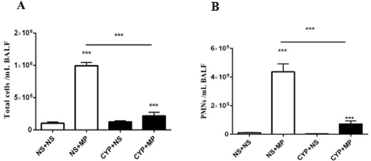
Figure 1.
Inflammatory cell recruitment into lung tissues after MP infection. The number of cells in BALF collected from mice pre-treated with CYP/saline and injected with 108 colony forming units of MP/saline for 96 h is shown. (A) Total number of cells in BALF. (B) PMNs in BALF. Data are presented as mean ± standard deviation (n = 5–7). MP, Mycoplasma pneumoniae; BALF, bronchoalveolar lavage fluid; CYP, cyclophosphamide; PMN, polymorphonuclear neutrophils; NS + NS, uninfected immunocompetent mice; CYP + NS, uninfected immunocompromised mice; NS + MP, infected immunocompetent mice; CYP + MP, infected immunocompromised mice. ***P < 0.001.

