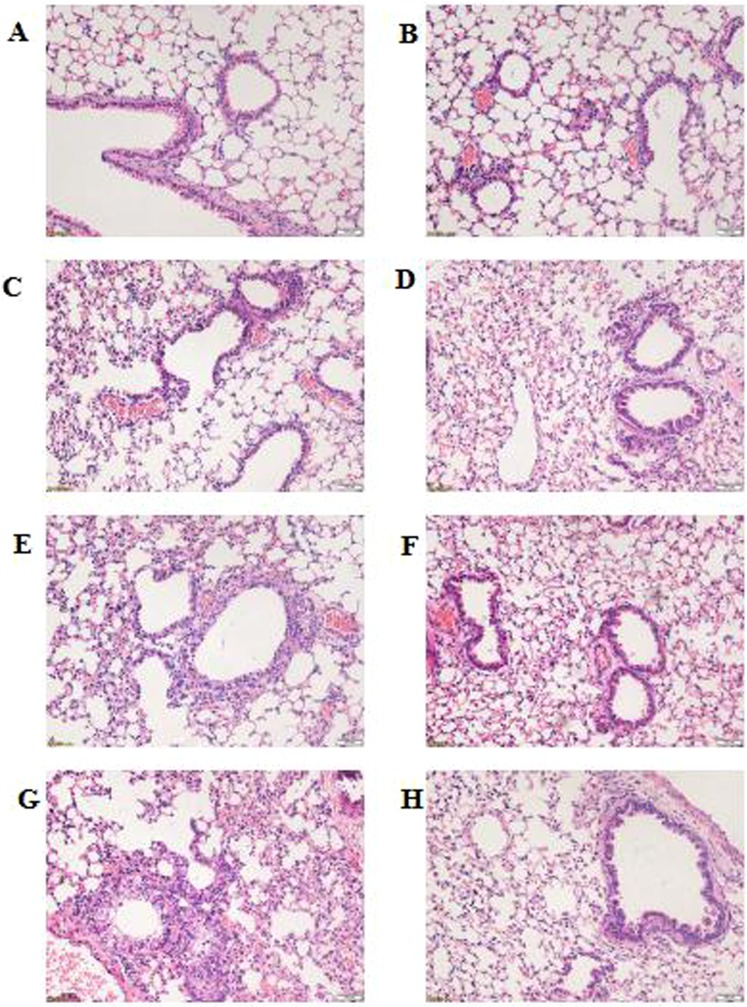
Figure 2.
Histopathological changes in lung tissues. Histological changes were determined at 0, 24, 48, and 96 h after Mycoplasma pneumoniae infection (hematoxylin–eosin staining; 200×). At 0 h: (A) NS + NS and (B) CYP + NS groups. At 24 h: (C) NS + MP and (D) CYP + MP groups. At 48 h: (E) NS + MP and (F) CYP + MP groups. At 96 h (G) NS + MP and (H) CYP + MP groups. NS + NS, uninfected immunocompetent mice; CYP + NS, uninfected immunocompromised mice; NS + MP, infected immunocompetent mice; CYP + MP, infected immunocompromised mice.

