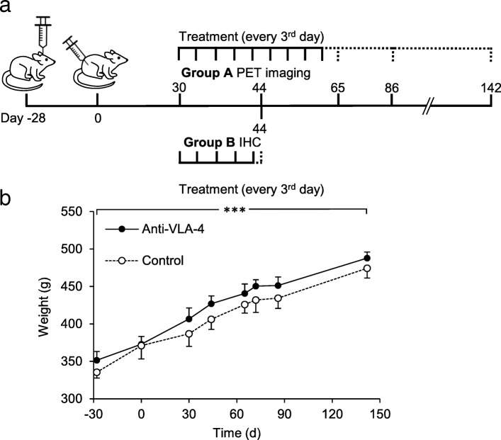
Fig. 1.
A schema of the experimental design showing the timeline of the procedures and changes in the rats’ body weight during the study. a Stereotaxic operation with BCG was performed on day − 28 (group A, n = 8 and group B, n = 4). Activation of the lesion was performed on day 0. Animals in group A were treated every third day with anti-VLA-4 mAb (n = 4) or with an isotype matched control mAb (n = 4) on days 30–61. The dashed line indicates the follow-up period after treatment. PET images were acquired on days 30, 44, 65, 86 and 142. Animals for IHC were treated every third day during days 30–44 and were killed on day 44 (group B). b Changes in the body weight of the animals during the PET imaging study. Results are shown as means (SD). ***p < 0.001; BCG Bacille Calmette-Guérin, IHC immunohistochemical, mAb monoclonal antibody, PET positron emission tomography

