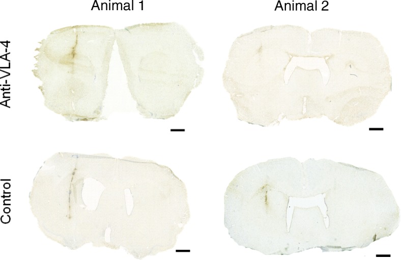
Fig. 4.

IHC staining with anti-Iba-1 of anti-VLA-4 mAb-treated and control animals after treatment over 14 days. Stereotaxic injection of BCG was performed on day − 28 (n = 4), and activation of the lesion was performed on day 0. Anti-VLA-4-treated animals were treated every third day on days 30–44 with anti-VLA-4 mAb (n = 2), and the control animals were treated with an isotype-matched control mAb (n = 2). The scale bar in each figure is 1500 μm. BCG Bacille Calmette-Guérin, Iba-1 ionised calcium-binding adapter molecule 1, IHC immunohistochemical, mAb monoclonal antibody, VLA-4 very late antigen-4
