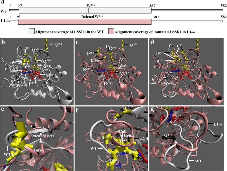
Fig. 9.
Predicted structure of LSSR1 in the WT and L1–4. The protein sequcence of LSSR1 in the WT and L1–4 were aligned with the same template c2zunB for structure prediction. a Alignment coverage of LSSR1 in the WT and L1–4. The three-dimensional (3D) model was constructed using these alignment covered sequences. b and c 3D model of the alignment covered sequence of LSSR1 in the WT and L1–4. d The merged structure in b and c. e-g Amplified image of the differential parts between the normal LSSR1 in the WT and the mutated LSSR1 in L1–4. White arrows point to residues (b, d) or domains (e-g) of the WT LSSR1, and black arrows point to residues (c, d) or domains (e-g) of the mutated LSSR1 in L1–4. Residues in yellow indicate the binding site, and in red indicate the catalytic site. The blue E205 (glutamine) may have both binding activity and catalytic activity, and the black W216 (tryptophan) is deleted in L1–4. E205 and E316 are the acid/base and the nucleophile, respectively

