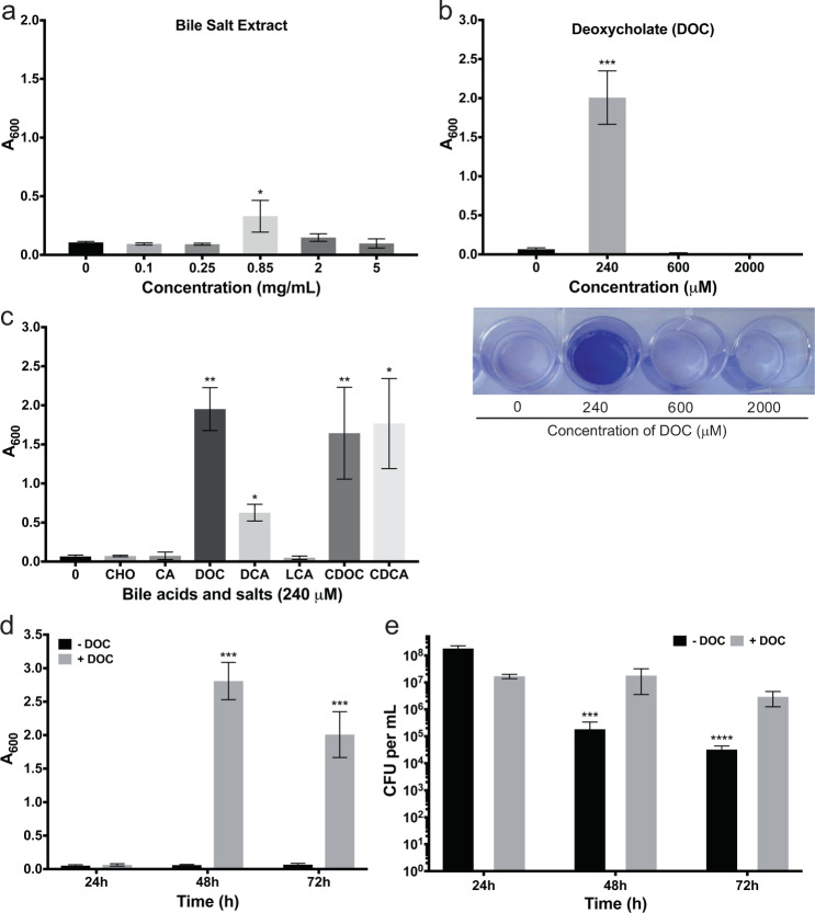
Fig. 1.
Effects of bile salts and deoxycholate on biofilm formation by C. difficile strain 630Δerm. a Bacteria were grown in BHISG supplemented with the indicated concentrations of bile salt extract or b deoxycholate. a, b Biofilm formation was evaluated at 72 h. In (b), representative image of a CV stained biofilm produced by cells cultured in the presence of increasing DOC concentrations. c Biofilm formation was evaluated after 48 h growth in the presence of 240 µM bile acids [cholic acid (CA), deoxycholic acid (DCA), lithocholic acid (LCA), chenodexycholic acid (CDCA)] or bile salts [cholate (CHO), deoxycholate (DOC) or chenodeoxycholate (CDOC)]. Asterisks indicate statistical significance determined with a Kruskal–Wallis test followed by an uncorrected Dunn’s test (*p ≤ 0.05, **p ≤ 0.001, ***p ≤ 0.001 vs BHISG with 0 mg/mL bile salts or 0 µM DOC). d Biofilm formation kinetics in the presence or absence of 240 µM DOC. Biofilm formation (a–d) was measured using a crystal violet assay that included two PBS washing before staining (see the Methods section). e Kinetics of CFU grown in the presence or absence of 240 µM DOC. The CFU counts were performed with unwashed biofilms (see the Methods section). Asterisks indicate statistical significance determined with a two-way ANOVA followed by a Fisher LSD test (***p ≤ 0.001, ****p ≤ 0.0001 vs 24 h). Data shown indicate the mean and the error bars represent the standard error of the mean of at least five experiments performed on different days

