Figure 7.
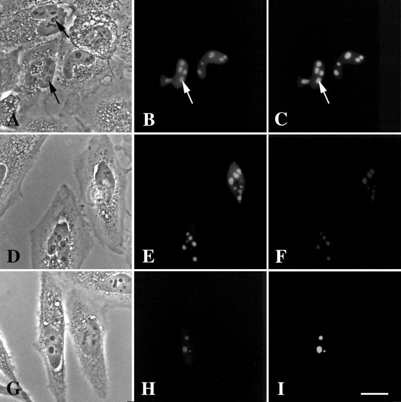
Coexpression of GFP-DmNopp140 and RFP-DmNopp140-RGG in transfected HeLa cells. (B, E, and H) Expression of GFP-DmNopp140. (C, F, and I) Expression of RFP-DmNopp140-RGG. (A–C) GFP-DmNopp140 and RFP-DmNopp140-RGG were expressed in approximately equal amounts based on fluorescence signals. Both proteins colocalized to the nucleoli. One nucleolus in a transfected cell appeared partially segregated by phase contrast microscopy (A, lower arrow), but no more so than a nucleolus in a nontransfected cell (A, upper arrow). (D–F) GFP-DmNopp140 (E) was overexpressed with respect to RFP-DmNopp140-RGG (F). Nucleoli appeared segregated by phase contrast microscopy (D), yet both proteins colocalized to the phase-light regions. (G–I) GFP-DmNopp140 (H) was underexpressed relative to RFP-DmNopp140-RGG (I). Nucleoli appeared morphologically normal by phase contrast microscopy (G), yet both proteins colocalized. Bar (A–I), 20 μm.

