Figure 2.
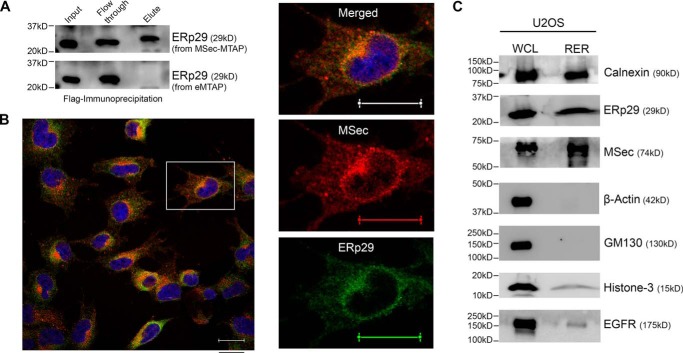
ERp29 interacts with MSec. A, FLAG immunoprecipitates of the MSec-MTAP and empty-MTAP U2OS cell lysates probed for ERp29 by immunoblotting. B, confocal images showing co-localization of MSec (red) with ERp29 (green) in the ER region. Pearson's correlation coefficient (Rcoloc) of the whole frame is 0.6766, and that of the selected cell is 0.6866. Scale bar, 20 μm. C, Western blotting of the whole-cell lysate (WCL) and rough endoplasmic reticulum (RER) fractions from U2OS cells probed for the respective proteins as indicated. Shown are ER-positive markers (calnexin and ERp29), cytosolic marker (β-actin), Golgi complex marker (GM130), nuclear marker (histone-3), and cell membrane marker (EGFR).

