Figure 3.
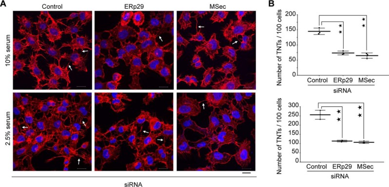
ERp29 is required for TNT formation. A, confocal microscopy images of cells co-stained with Alexa FluorTM 594–conjugated phalloidin (red, to visualize F-actin) and 4′,6-diamidino-2-phenylindole (blue, to visualize the nucleus) after 48 h of the indicated siRNA transfection. Arrows, TNTs connecting neighboring cells. Scale bar, 20 μm. B, quantification of the number of TNTs per 100 cells from the confocal images in both normal (10% serum) and stress (2.5% serum) conditions, respectively. Data represent mean ± S.D. (error bars) based on three independent experiments, 100 cells counted per experiment (paired t test, two-tailed; **, p < 0.01).

