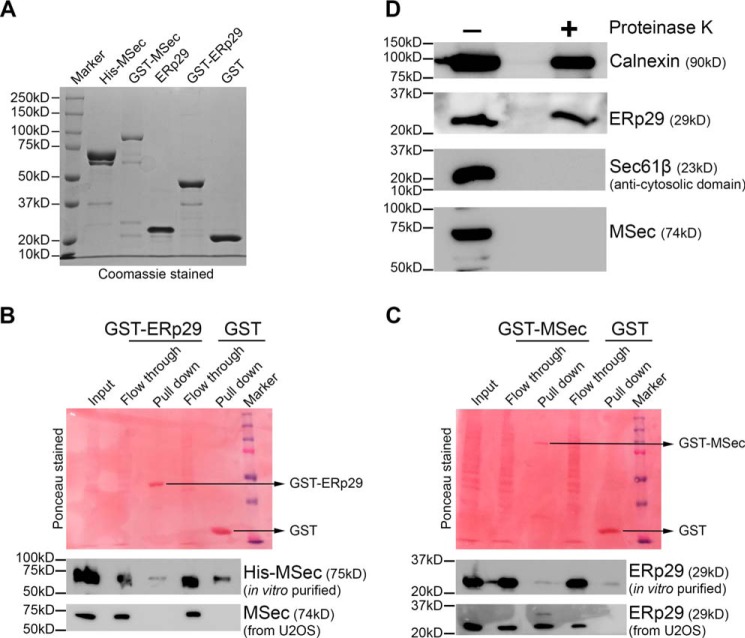
Figure 7.
Characterization of the ERp29–MSec interaction. A, SDS-PAGE profiles (Coomassie-stained) of the purified reombinant MSec and ERp29 proteins as indicated. B, purified ERp29 shows no interaction with either purified MSec or MSec from cell lysate. The bottom strips show immunoblots with the respective antibodies as indicated. C, purified MSec does not interact with purified ERp29 but interacts with ERp29 from cell lysate. The bottom strips show immunoblots with the respective antibodies as indicated. D, protease protection assay with proteinase K reveals that MSec is associated with the ER surface toward its cytosolic side. Markers used in immunoblots were calnexin (ER lumen) and Sec61β-cytosolic side (ER surface).

