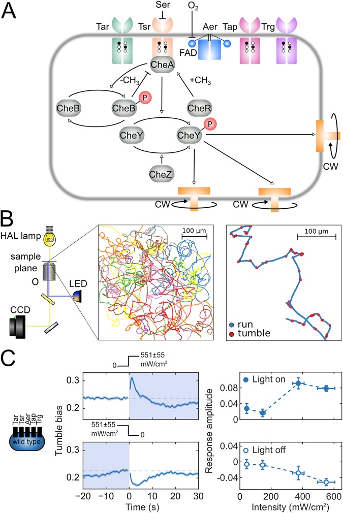
FIG 1.
Phototaxis in E. coli. (A) Schematic of the chemotaxis network. Five types of chemoreceptors (Tar, Tsr, Aer, Trg, and Tap), sensitive to different extracellular and intracellular cues, modulate the activity of the kinase CheA, which phosphorylates the signaling molecule CheY. Phosphorylated CheY binds to flagellar motors, causing them to switch to CW rotation. Chemotactic adaptation is mediated by the methyltransferase CheR and the methylesterase CheB. Methylation of receptor sites (black circles) by CheR increases kinase activity. CheB, when phosphorylated by active CheA, demethylates receptors (white circles) and decreases kinase activity. (B) Experimental and data analysis framework for studying phototaxis in E. coli. An inverted light microscope with a 20× objective (O) and a wide-range halogen (HAL) lamp (yellow light path) images swimming cells, and a blue LED (blue light path) stimulates cells. Movies of swimming bacteria are captured by a CCD camera. Bacteria are detected in each movie frame and their coordinates are linked into trajectories, which are filtered and analyzed to assign runs and tumbles. (C, left and center) Response of the wild-type strain RP437 (schematic indicates that all receptor types are present) to a turn on and turn off of blue light with an intensity of 551 ± 55 mW/cm2. Light exposure is indicated by shading and by the light intensity profiles above the plots. Each point on the time trace is the average of the tumble biases of ∼6,000 to 7,000 trajectories. The shading around the time trace represents the standard error of the mean tumble bias. (C, right) Amplitude of the responses to turn on (top) and turn off (bottom) as a function of light intensity.

