
FIG 4.
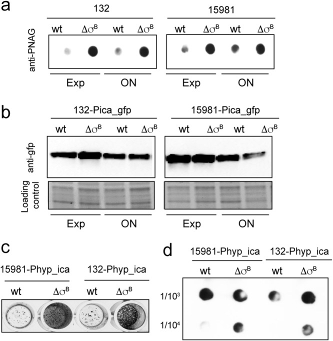
Analysis of ica operon expression in σB mutants. (a) PNAG accumulation at different growth stages: exponential (Exp) and stationary (ON). Samples diluted 1:100 or 1:5,000 were spotted onto nitrocellulose membranes. PNAG production was detected with anti-PNAG antiserum. (b) Effects of σB deletion on ica promoter activity in S. aureus strains 15981 and 132 (wt) and their respective ΔσB mutants carrying pCN52-Pica_gfp at exponential and stationary phases. Expression of GFP under the control of the ica promoter (Pica_gfp) was determined by Western blotting using monoclonal antibodies. (c) Biofilm formation by S. aureus 15981 Phyp_ica and 132 Phyp_ica and their respective σB mutants on polystyrene microtiter plates after 4 h of incubation. The bacterial cells were stained with crystal violet. (d) PNAG accumulation in cell extracts of S. aureus 15981 Phyp_ica and 132 Phyp_ica and their respective σB mutants. PNAG production was detected by dot blot analysis using anti-S. aureus PNAG antiserum.