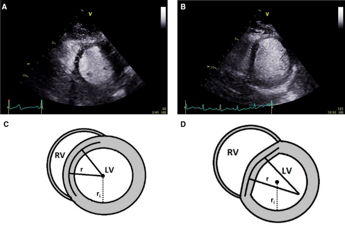
Figure 1.

Septal flattening from diastolic ventricular interaction causes a D‐shaped left ventricle, and can be quantified from the normalized radius of curvature. A, A resting contrast echocardiogram in the parasternal short axis view of the left ventricle of a patient with heart failure with preserved ejection fraction, demonstrating a circular interventricular septum at end‐diastole. B, In the same patient during exercise, the subsequent development of pulmonary hypertension leads to pericardial constraint and diastolic ventricular interaction, flattening the septum and resulting in a D‐shaped LV. C, The shape of the left ventricular short axis image is calculated from the ratio of the radius extrapolated from the demarcated septum (r) to the cavity radius (r i), called the normalized radius of curvature. This is close to 1 at rest. D, During exercise in patients with diastolic ventricular interaction, the flattened septum produces a calculated NRC value much >1. LV indicates left ventricle; NRC, normalized radius of curvature; RV, right ventricle.
