
Figure 1.
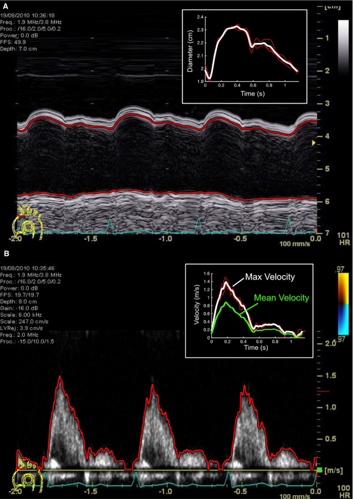
M‐mode echocardiographic still‐image of aortic arch diameter (A) and blood velocity (B), with automated edge segmentation and average beat files displayed in the inset boxes.
Official websites use .gov
A
.gov website belongs to an official
government organization in the United States.
Secure .gov websites use HTTPS
A lock (
) or https:// means you've safely
connected to the .gov website. Share sensitive
information only on official, secure websites.
M‐mode echocardiographic still‐image of aortic arch diameter (A) and blood velocity (B), with automated edge segmentation and average beat files displayed in the inset boxes.