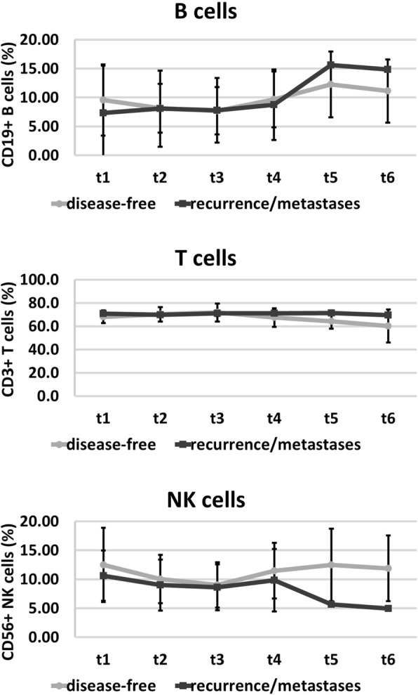
Fig. 3.

Immunophenotyping of the percentages of major lymphocyte subpopulations such as CD3−/CD56+ NK cells, CD3+ T cells, CD19+ B cells before (t1), after 30 Gy (t2), at the end of RT (t3), and in the follow-up period (t4, 6 weeks after RT; t5, 6 months after RT; t6, 1 year after RT) in disease-free patients (closed circles) and patients who developed recurrence or distant metastases (closed squares). The data show mean values ± standard deviation of the percentage of positively stained cells
