. 2019 Jan 3;48(18):5978–5986. doi: 10.1039/c8dt04294b

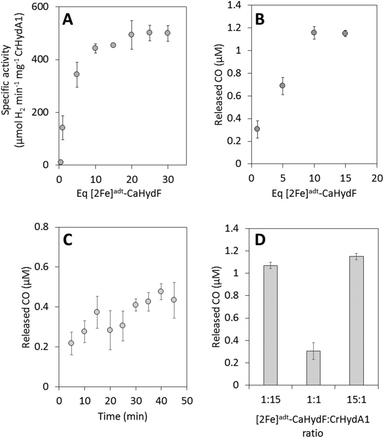
Fig. 3. The activation of apo-CrHydA1 using [2Fe]adt-CaHydF probed by enzymatic assays and CO release. (A) H2 production assay, CrHydA1 titration curve: 0.04 μM apo-CrHydA1 (1 equivalent) was incubated with 0.04–1.2 μM of [2Fe]adt-CaHydF in 100 mM K-phosphate pH 6.8 buffer at 37 °C for 60 minutes. After 60 minutes, hydrogenase activity was determined via a methyl viologen assay. (B) CO release, CrHydA1 titration curve: 2 μM CrHydA1 (1 equivalent) was incubated with 2–30 μM of [2Fe]adt-CaHydF for 25 minutes. (C) Time dependence of CO release: 2 μM apo-CrHydA (1 equivalent) was incubated with 2 μM [2Fe]adt-CaHydF (1 equivalent) for 5–45 min. (D) CO release as a function of the relative ratio between apo-CrHydA1 and [2Fe]adt-CaHydF – 2 μM [2Fe]adt-CaHydF to 30 μM apo-CrHydA1 (1 : 15), 2 μM [2Fe]adt-CaHydF to 2 μM apo-CrHydA1 (1 : 1) and 30 μM [2Fe]adt-CaHydF to 2 μM CrHydA1. All CO release experiments were performed at room temperature in 100 mM Tris-HCl pH 7.0 300 mM KCl.

Fig. 3