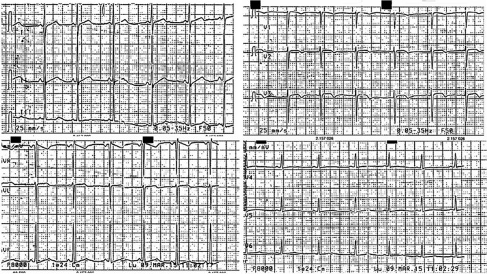
Figure 1.

Electrocardiogram showing sinus rhythm with alterations of the ventricular repolarization as inverted T wave in V2‐V3, poor progression of the r wave in the precordial leads and high voltages in the peripheral leads

Electrocardiogram showing sinus rhythm with alterations of the ventricular repolarization as inverted T wave in V2‐V3, poor progression of the r wave in the precordial leads and high voltages in the peripheral leads