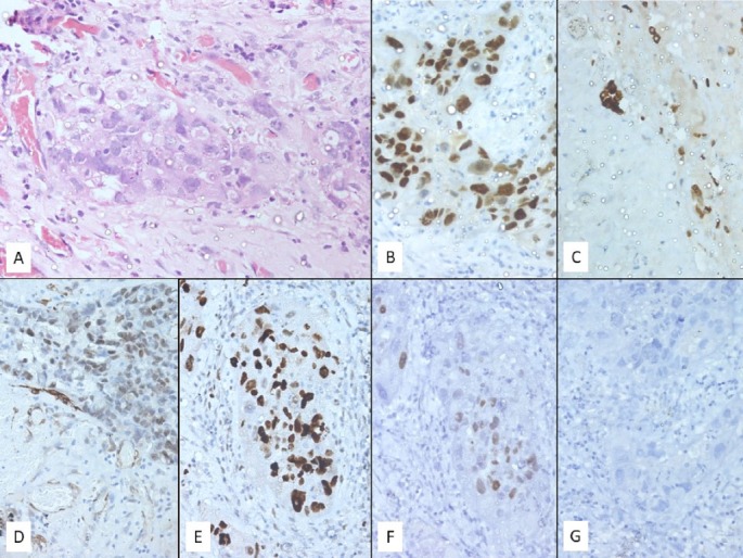
Figure 4.

H&E stained section (A) (400× magnification) showing atypical cell clusters in the pericardial wall. These cells show marked nuclear pleomorphism with coarse chromatin, prominent nucleoli and scant pale eosinophilic cytoplasm. On immunocytochemistry these cells are positive for antibodies against PAX8 (B) (intense and nuclear), cytokeratin 7 (C) (intense and cytoplasmic), WT1 (D) (focal and nuclear), p53 (E) (intense and nuclear) and ER (F) (pale and nuclear) while being negative for PR (G).
