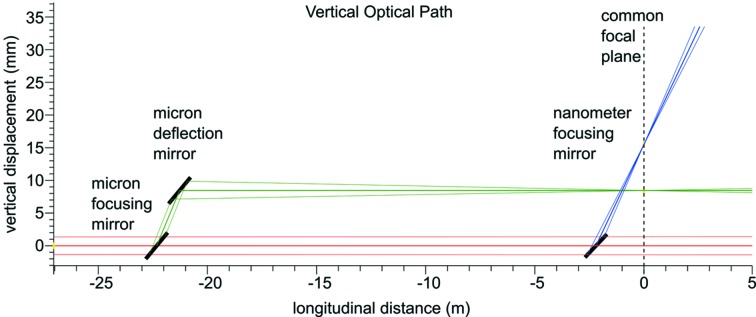
Figure 6.
Representation of the vertical optical layout of the SPB/SFX instrument. The incident beam from XTD9 is shown in red, with the 1 µm-scale system in green and the 100 nm-scale system in blue. The 0 m mark in the longitudinal distance denotes the common focal plane of the two systems. Figure adapted from Bean et al. (2016 ▸).

