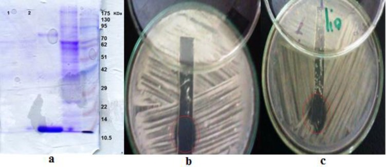
Figure 2.
Gel electrophoresis analysis of antifungal peptides derived of Bacillus strains submitted to SDS-PAGE and stained for proteins with Coomassie blue (a) antifungal peptide derived from Bacillus strains from wells 1 and 2, and protein marker from well 5 were loaded on the gel. The peptide derived from B.pumilus ZED17 (b) B.pumilus DFAR8 (c) in unstained gel tested for antifungal activity on R. solani. In images b and c, the visible inhibition zones were observed in the cycles

