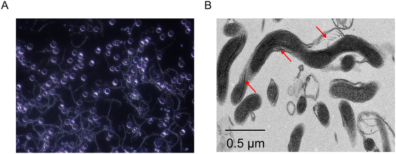
Figure 1.
The unique structure of spirochetes. Panel A presents a dark-field microscopic image of Borrelia hermsii, a relapsing fever spirochete, in blood collected from an infected mouse. Mice were infected by needle inoculation and blood samples were collected 3 days post-inoculation. The characteristic spiral morphology shared by all spirochetes is readily visible. Note, there is high density spirochetemia. In contrast to relapsing fever, spirochetemias are not common in mammals infected with Lyme disease. Panel B presents a transmission electron micrograph of the oral spirochete Treponema denticola. The endoflagella bundles that are unique to spirochetes are indicated by the arrows.

