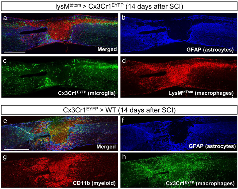
Figure 1. Bone marrow chimeras reveal distinct spatial distribution of macrophages and microglia at the spinal cord injury site.
(a-d) LysM-Cre mice were bred to Rosa26-tdTomato mice to generate LysMtdTom reporter mice in which myeloid cells are labeled with tdTomato. Bone marrow from LysMtdTom donor mice were transplanted into irradiated Cx3Cr1EYFP recipient mice to generate LysMtdTom > Cx3Cr1EYFP chimera mice in which microglia are labeled with EYFP and hematogenous macrophages are labeled with tdTomato. At 14 days after SCI, microglia (c) are distributed mostly around in the lesions site in the GFAP+ astroglial scar region (b), whereas macrophages are located both within the GFAP− lesion site and in the surrounding astroglial scar (d). (e-h) Bone marrow from Cx3Cr1EYFP donor mice were transplanted into irradiated wildtype recipient mice to generate Cx3Cr1EYFP > WT chimera mice in which only macrophages are labeled with EYFP. In theory, this should result in a similar distribution pattern as lysMtdTom macrophages shown in (d). However, large areas of the GFAP− region (f) are devoid of EYFP signal (h) even though large number of CD11b+ myeloid cells can be observed immunohistochemically (g). This leads to the false impression that macrophages are located in the surrounding astroglial scar region, and that the CD11b+/EYFP− cells in the GFAP− region are microglia. Scale bar in A, E = 500μm. Unpublished images from [107].

