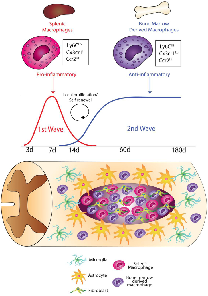
Figure 2: Schematic illustration of putative sources of macrophages after spinal cord injury.
Pro-inflammatory splenic macrophages comprise the major source of the initial wave of macrophage influx. A second wave of anti-inflammatory macrophages could be from either the bone marrow or from a self-renewing source at the injury site. The spinal cord injury site is comprised of a fibrotic core comprised of non-neural cells such as fibroblasts and macrophages surrounded by neural cells such as astrocytes and microglia. Bone marrow chimera studies (Figure 1) demonstrate the presence of Cx3cr1lo macrophages in the fibrotic region, and Cx3cr1hi macrophages in both the fibrotic and surrounding neural tissue. These two types of macrophages could correspond to the pro-inflammatory and anti-inflammatory subtypes from the spleen and bone marrow, respectively.

