
FIGURE 2.
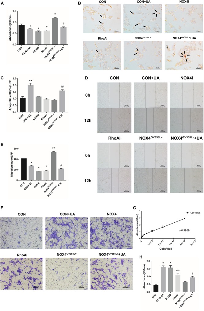
The regulatory effects of NOX4 and RhoA as well as the UA intervention effects on the migration and invasion of HSCs after TGF-β1 treatment (10 ng/mL) for 48 h. (A) Cell proliferation detected by the MTS assay. (B,C) Cell apoptosis levels detected by the TUNEL assay. Black arrows: typical apoptotic cells. Original magnification, 200×. Percentage of apoptotic cells = apoptotic cells/cell count per high-power field; 10 different fields of vision were randomly selected in each group. (D,E) Cell scratch assay. Original magnification, 100×. (F,H) Cell invasion detected by the Transwell invasion system. Original magnification, 200×; (G) MTS indirect cell count curve. r = 0.9959. The data are presented as the means ± SEMs of three replicates. ∗p < 0.05, ∗∗p < 0.01 versus the CON group; #p < 0.05, ##p < 0.01 versus the NOX4GV 358Lv group. ∆p < 0.05 versus the NOX4i group.