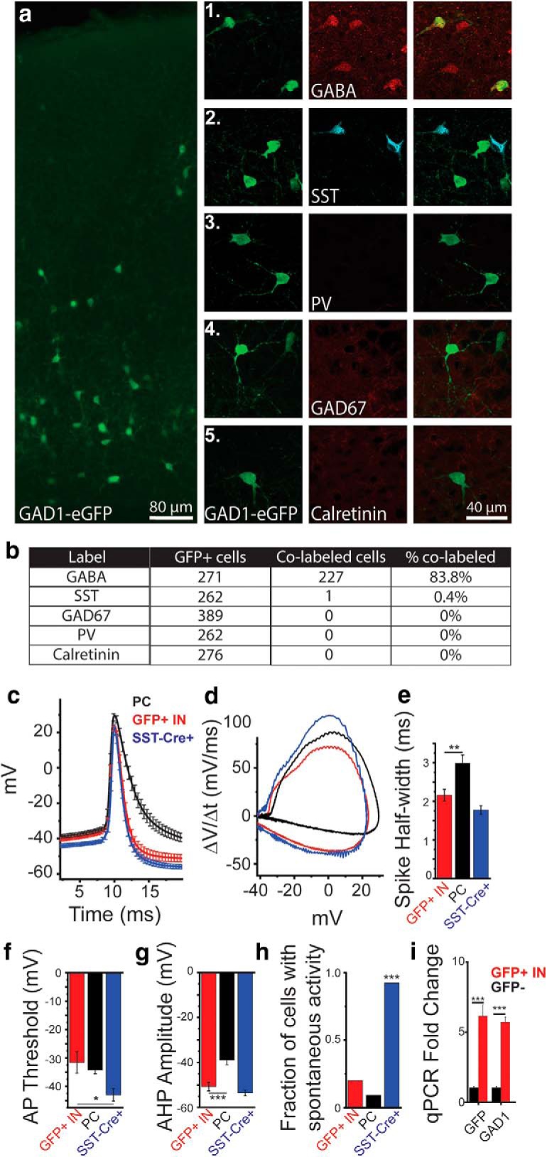
Figure 4.

G42 mice identify GABAergic non-SST INs at P9. a, Immunohistochemical labeling of GFP in the somatosensory cortex (SSC) of G42 (GAD1-eGFP) mice at P9. Coimmunolabeling of GFP in G42 mice with markers on inhibitory INs. a1–a5, GFP (left). a1, GABA. a2, Somatostatin. a3, PV. a4, GAD67. a5, Calretinin (middle), colabel (right) in layer V of the SSC. b, Table showing abundant colabeling of GFP+ cells with GABA immunolabeling at P9. Sections from 4 mice were used to quantify colocalization. c, Average evoked APs from whole-cell recordings from GFP+ cells in G42 mice (red), L5Ps (black), and Td-tomato+ neurons in SST-Cre X Ai9 mice (blue). d, dV/dt analysis of APs shown in c. e, AP half-width. f, AP threshold. g, Afterhyperpolarization amplitude. h, Fraction of cells showing spontaneous AP firing in GFP+ cells in G42 mice (red), L5P neurons (black), and Td-Tomato+ neurons in SST-Cre X Ai9 mice (blue). *p < 0.05 (one-way ANOVA). **p < 0.01 (one-way ANOVA). ***p < 0.001 (one-way ANOVA). i, qPCR analysis of GFP+ and GFP− cells at P9 in G42 mice separated via FACS. ***p < 0.001 (paired t test).
