Figure 2.
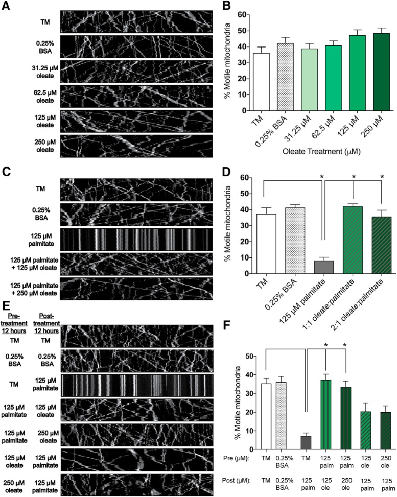
MUFA treatment preserves axonal mitochondrial motility in cultured DRG neurons. A, Kymograph analysis of DRG axons treated for 24 h with TM, vehicle only (0.25% BSA), and varying concentrations (31.25–250 μm) of oleate. B, Percentage of motile mitochondria as observed in A. C, Kymograph analysis of DRG axons treated for 24 h with TM, vehicle only (0.25% BSA), 125 μm palmitate alone, and 125 μm palmitate with 125 or 250 μm oleate. D, Percentage of motile mitochondria as observed in C. E, Kymograph analysis of DRG neurons treated for 24 h with TM, vehicle alone (0.25% BSA), pre-treatment (12 h) with 125 μm palmitate followed by post-treatment (12 h) with 125 or 250 μm oleate, or pre-treatment (12 h) with 125 or 250 μm oleate followed by post-treatment (12 h) with 125 μm palmitate. F, Percentage of motile mitochondria as observed in E. All data represent n = 16–23 neurons per condition (A, B), n = 18–23 neurons per condition (C, D), and 12–16 neurons per condition (E, F): one-way ANOVA with Tukey's multiple-comparisons test. *p < 0.0001.

