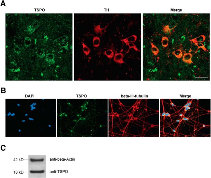
Figure 7.
TSPO expression in dopaminergic neurons. A, Immunofluorescence staining of 8-week-old WT control mice (substantia nigra). Sections were stained with antibodies against TSPO (left, green) and TH (middle, red). Right, Merged image. Scale bar, 20 μm. B, Immunofluorescence staining of LUHMES cells. Left, DAPI (blue), antibody against TSPO (green), antibody against β-III-tubulin (red). Right, Merged image. Scale bar, 50 μm. C, Western blot analysis of TSPO expression in LUHMES cells.

