Figure 2.
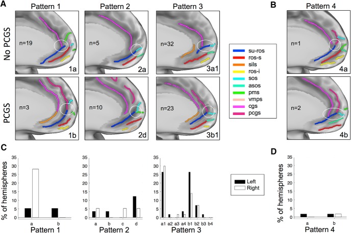
vmPFC anatomical patterns. A, Examples of the three common types of sulcal patterns in the vmPFC. The patterns depend on how the SU-ROS merges with other sulci (white circles) and the absence (first row) or presence (second row) of a PCGS. In type 1, the SU-ROS merges with either the CGS (1a) or the PCGS (1b). In type 2, the SU-ROS merges with one or more polar/supraorbital sulci but not with the CGS (2a) or the PCGS (2d). In type 3, the SU-ROS merges with both polar/supraorbital sulcus and CGS (3a1) or PCGS (3b1). B, In the rare type 4, the SU-ROS merges with both CGS and PCGS, without a merging (4a) or with (4b) a merging with a polar/supraorbital sulcus. n indicates the number of hemispheres exhibiting each pattern. C, D, Percentage of left (black) and right (white) hemispheres displaying each pattern and subtype of patterns. 1a, only CGS (left, 5.3%; right, 28.1%); 1b, only PCGS (left, 5.3%; right, 0%); 2a, only SOS (left, 3.5%; right, 5.3%); 2b, only ASOS (left, 3.5%; right, 0%); 2c, only PMS (left, 0%; right, 5.3%); 2d, two polar/supraorbital sulci (left, 12.3%; right, 5.3%); 3a, with CGS and with SOS (3a1: left, 26.3%; right, 29.8%), or ASOS (3a2: left, 1.8%; right, 0%), or PMS (3a3: left, 0%; right, 1.8%), or with two polar/supraorbital sulci (3a4: left, 3.5%; right, 1.8%); 3b, with PCGS and with SOS (3b1: left, 26.3%; right, 14.0%), or ASOS (3b2: left, 7.0%; right, 7.0%), or PMS (3b3: left, 1.8%; right, 0%), or two polar/supraorbital sulci (3b4: left, 0%; right, 0%); 4a, CGS and PCGS (left, 1.8%; right, 0%); and 4b, CGS, PCGS, and polar/supraorbital sulcus (left, 1.8%; right, 1.8%).

