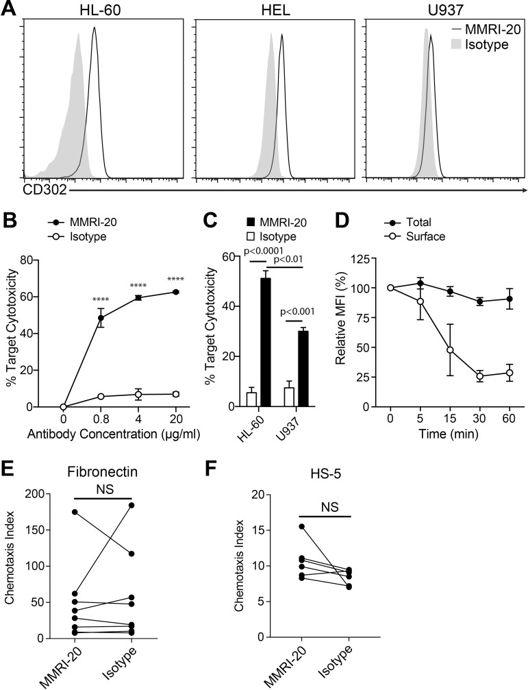
Fig 2. Antibodies targeting CD302 are able to be internalised and mediate ADCC of target cells.
(A) Flow cytometry histograms showing the surface expression of CD302 on leukemic target cell lines as determined by staining with the MMRI-20 compared to a mouse IgG1 isotype control. (B) CD302 internalisation by HL-60 cells determined by flow cytometry. Total staining determined by MMRI-20-FITC 37°C incubation for the indicated times after which residual surface CD302 was measured with anti-mouse IgG-PE at 4°C. MFI of antibody staining is reported relative to pre-incubation levels. (C) MMRI-20 induced ADCC against HL-60 target cells. Calcein-AM labelled HL-60 were incubated for 18h with mouse spleen effectors at a 1:10 ratio, together with1000U IL-2 and the indicated concentrations of MMRI-20 or isotype control mAb. Target cell killing was measured as 7-AAD+ Calcein-AM+ cells by flow cytometry and presented relative to death in target alone (0%) or with 2% Triton X solution (100%). **** p<0.0001, two-way ANOVA. (D) ADCC elicited against HL-60 (CD302hi) and U937 (CD302lo) leukemic targets using 20μg/ml MMRI-20 or isotype mAb control. Experiments representative of three experiments. Differences tested by two-way ANOVA. (E-F) HL-60 cells were incubated with either MMRI-20 or isotype control mAb for 30 mins at 37°C and tested for their ability to migrate across 5 μm transwells coated with (E) fibronectin or (F) HS-5 cells towards 160 ng/ml CXCL12 or media alone. Cells in bottom chamber were enumerated after 4h by using flow cytometry and migration presented as the chemotaxis index. Circles connected by lines represent individual paired experiments. No significant difference (NS) between MMRI-20 and isotype group (paired t-test).

