. 2019 May 10;8:e44436. doi: 10.7554/eLife.44436

Figure 1. Murine CypA has a diminished ability to facilitate HCV replication.

(A) An amino acid sequence alignment of CypA from diverse species. Similar amino acids are indicated in boxed, bold capital letters while differences are lowercase. Species are arranged from top to bottom in increasing evolutionary distance from human. For pigtailed macaque, all experiments utilized a TRIM5-CypA fusion – only the residues of the CypA portion of the fusion are depicted here. (B) Huh7.5 cells expressing an shRNA against endogenous human CypA (Huh7.5-shRNA CypA) were transduced to express different CypA orthologs and then infected with a HCV reporter genome expressing secreted Gaussia luciferase (Jc1-Gluc, MOI = 0.1). At five dpi, the luciferase activity of the supernatants was assessed as a proxy for viral replication. CypA rescue efficiency is shown normalized to Huh7.5-shRNA CypA transduced with human CypA, which is 100% identical at the amino acid level to chimpanzee, bonobo, gorilla, olive baboon and rhesus macaque CypA. Results shown are from two representative experiments, each with triplicate samples. Lines and error bars represent the mean ± SD. Ordinary two-way ANOVA test performed followed by Dunnett’s multiple comparisons test with all means compared to that of the +human CypA line. Chimp/Ch, chimpanzee; Bo, bonobo; Go, gorilla; Orang, orangutan; Rhesus mac/RhM, rhesus macaque; Pt mac, pigtailed macaque; Olive bab/Bab, olive baboon; Sq monkey, squirrel monkey. ****, p<0.0001; ns, not significant.

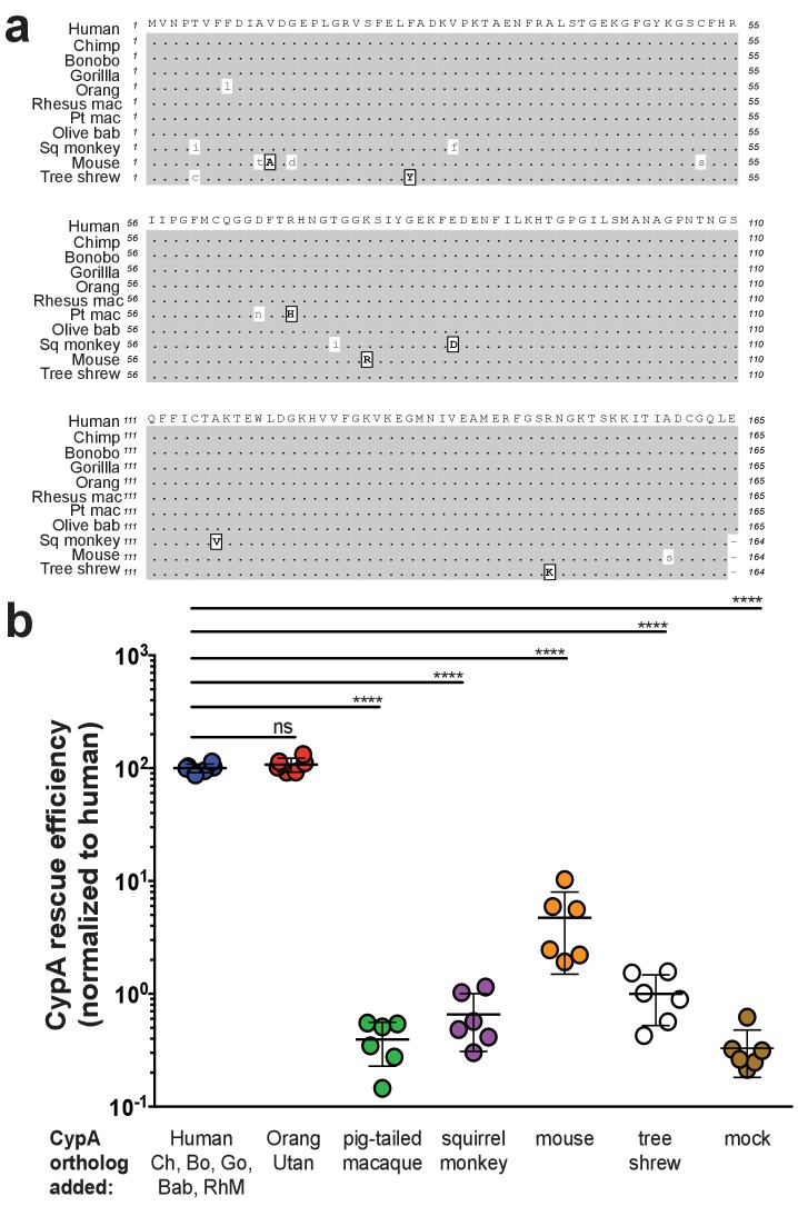
Figure 1.

Figure 1—figure supplement 1. Assessment of Huh7.5-shRNA CypA cells.

Figure 1—figure supplement 1.

(A) Western blot demonstrating knockdown of human CypA in Huh7.5-shRNA CypA cells relative to the parental Huh7.5 cells and those expressing an shRNA against an irrelevant target (Huh7.5-shRNA irrel). Quantification of signal intensity was done using LI-COR Image Studio Software (version 4.0) with background detection set to ‘Median.’ CypA signal intensity was normalized based off that of β-actin and is expressed here relative to Huh7.5 cells (set at 100%). (B) HCV replication is specifically diminished in Huh7.5-shRNA CypA cells and not Huh7.5-shRNA irrel cells. Infection performed with Jc1-Gluc (MOI = 0.1). On even-numbered days, cells were washed with PBS and the media changed to assess de novo replication. Lines and error bars represent the mean ± SD.
Figure 1—figure supplement 2. Transduction efficiency of CypA orthologs in Huh7.5-shRNA CypA cells.

Figure 1—figure supplement 2.

Bicistronic constructs expressing the CypA orthologs of interest followed by an IRES-regulated eGFP-ubiquitin-neomycin fusion protein were used to transduce Huh7.5-shRNA CypA cells. Transduction efficiency assessed via flow cytometry and representative data are summarized here from one set of transductions (A). Cell lysates were collected from Huh7.5-shRNA CypA cells transduced with these various constructs and expression determined by western blot (please see Supplementary file 1). Quantification of CypA expression was determined by taking the signal from the CypA band divided by the signal from the β-actin band and then this value divided by that from the untransduced Huh7.5-shRNA CypA cells to give the relative expression (B). Signal intensity was determined using LI-COR Image Studio Software (version 4.0) with background detection set to ‘Lane.’ The ‘a’ and ‘b’ above each bar delineate the different pairs of antibodies used to assess CypA expression. For ‘a,’ the signal was quantified from rabbit anti-CypA and mouse anti-β-actin; for ‘b,’ the signal was quantified from mouse anti CypA and rabbit anti- β-actin. ND, non-detected; Mn, pigtailed macaque.