. 2019 May 10;8:e44436. doi: 10.7554/eLife.44436

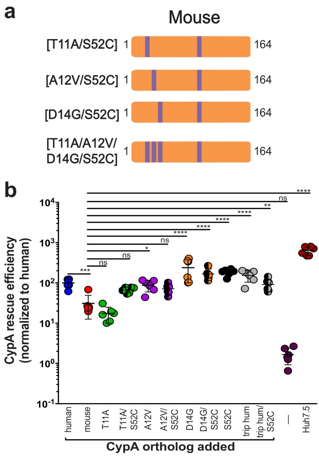
Figure 3. Humanizing residue 52 in the murine CypA mutant T11A/A12V/D14G does not further increase rescue efficiency.

(A) Schematic of the additional humanized mouse CypA mutants tested. (B) Huh7.5-shRNA CypA cells were transduced with the mutants shown in (A) and infected with Jc1-Gluc at MOI = 0.1. Supernatants were assessed for Gaussia luciferase activity as a proxy for HCV replication at five dpi, and the rescue efficiency of each mutant was normalized to Huh7.5-shRNA CypA transduced with human CypA. Results shown are from two representative experiments, each with triplicate samples. Lines and error bars represent the mean ± SD. Ordinary two-way ANOVA test performed followed by Sidak’s multiple comparison test, with all means compared to that of the +mouse CypA line. *, p<0.05; **, p<0.01; ***, p<0.001; ****, p<0.0001, ns, not significant.

Figure 3.

Figure 3—figure supplement 1. Transduction efficiency of additional mouse-S52C CypA mutants in Huh7.5-shRNA CypA cells.

Figure 3—figure supplement 1.

Bicistronic constructs expressing the CypA mutants shown in Figure 3A followed by an IRES-regulated eGFP-ubiquitin-neomycin fusion protein were used to transduce Huh7.5-shRNA CypA cells. Transduction efficiency assessed via flow cytometry and representative data are summarized here from one set of transductions (A). Cell lysates were collected from Huh7.5-shRNA CypA cells transduced with these various constructs and expression determined by western blot (please see Supplementary file 1). Quantification of CypA expression was determined by taking the signal from the CypA band divided by the signal from the β-actin band and then this value divided by that from the untransduced Huh7.5-shRNA CypA cells to give the relative expression (B). Signal intensity was determined using LI-COR Image Studio Software (version 4.0) with background detection set to ‘Lane.’ The ‘a’ and ‘b’ above each bar delineate the different pairs of antibodies used to assess CypA expression. For ‘a,’ the signal was quantified from rabbit anti-CypA and mouse anti-β-actin; for ‘b,’ the signal was quantified from mouse anti CypA and rabbit anti- β-actin.